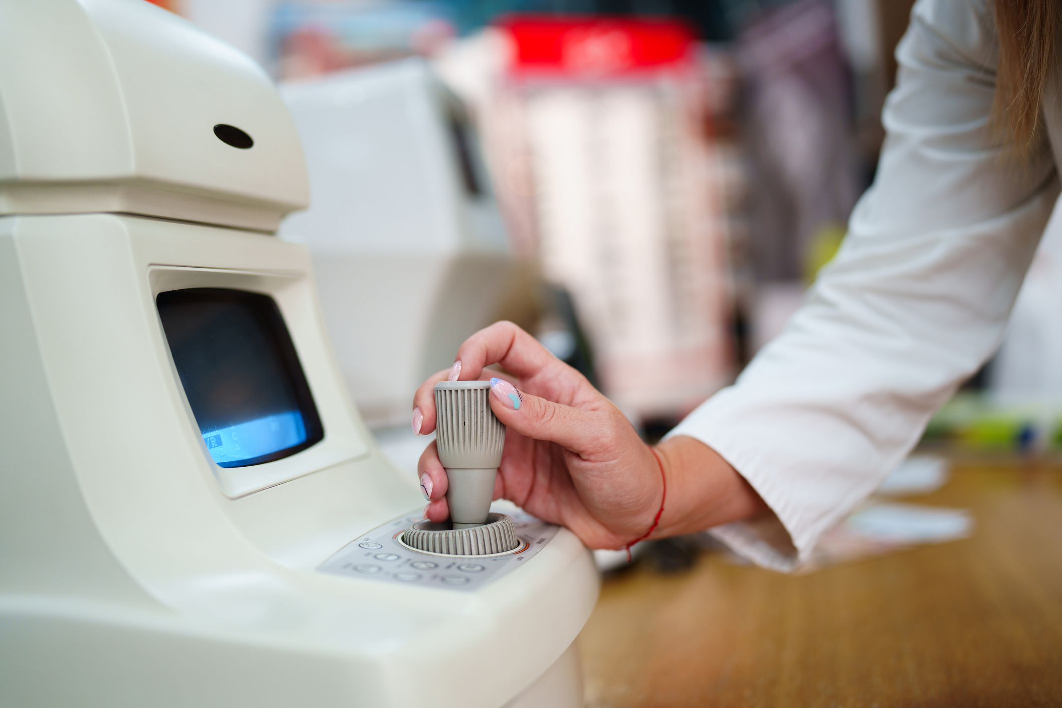
Optometrists are encouraged to adopt the World Council of Optometry’s (WCO) Standard of Care for Myopia Management, which outlines three core components: mitigation, measurement, and management of childhood myopia.1 While specialized equipment is not essential to begin providing myopia management, it can greatly enhance the quality and effectiveness of care. With myopia prevalence rates increasing globally2 and in Canada3 and myopia management becoming an increasingly core component of practice, investing in tools such as axial length measurement devices, corneal topographers, and other diagnostic technologies may be a worthwhile consideration. This article will go through the different instruments that may enhance myopia management in practice.
Instruments for Axial Length Measurement
The main objective of myopia control is to slow down the eye’s axial growth. While measuring axial length isn’t a requirement to provide myopia management, it offers valuable insights into a child’s risk of developing myopia4,5 and their long-term eye health outlook.6 This is particularly important in orthokeratology, where the refractive error is intentionally altered—making axial length a more reliable way to assess progression. These measurements can also guide clinical decisions; for instance, if a child presents with low myopia but has a longer-than-expected axial length, this could indicate a higher risk of future complications and may justify a more proactive treatment plan.
Optical biometers measure axial length using various technologies; however, these technologies differ in precision, repeatability, and ease of use—factors especially important for effective myopia management. Two main technologies used are interferometry (optical biometry) and ultrasound:
- Interferometry (optical biometry) is a non-contact optical method that uses light waves to measure the eye. Devices like the IOLMaster, Lenstar, and Pentacam AXL use this technique to provide highly accurate and repeatable measurements.7-11 These instruments are quick, comfortable for children, and ideal for tracking even subtle axial elongation.
- Ultrasound biometry, or A-scan, uses sound waves and requires a probe to touch the eye after anaesthetic drops are applied. It has been used in earlier myopia studies12-15 but is less accurate and more dependent on the skill of the operator. The pressure applied during the test can affect the results, and it’s less comfortable for children.
Overall, interferometry is preferred due to its higher precision (detecting changes as small as 0.03D, compared to 0.30D with ultrasound – the latter being no better than refraction to track myopia changes), non-contact method, and better suitability for monitoring myopia over time. To understand the differences between the two methods of axial length measurement in more detail, you can read the article Choosing an instrument to measure axial length on MyopiaProfile.com. To understand how to use axial length measurements in practice, you can read the article How much axial length growth is normal?
Instruments for Corneal Assessment
A variety of corneal imaging instruments are available to support myopia management and contact lens practice, including the Medmont E300, Keratron Scout (Optikon), Tomey TMS Series Corneal Topographers, OCULUS Keratograph 5M, and the Nidek OPD-Scan III.
Corneal topography, commonly found in these devices, uses Placido disc technology to map the anterior corneal surface, providing useful information about its shape, curvature, and eccentricity. While not essential for every case, it can assist with more precise fitting of orthokeratology and other specialty lenses and may add value to contact lens assessments. In myopia management, topography is commonly used to support orthokeratology lens design and to monitor lens centration and corneal changes over time.
Tomography, which uses Scheimpflug imaging to capture both the anterior and posterior corneal surfaces, offers additional detail. This can be helpful in detecting irregularities such as keratoconus or unexplained visual symptoms. While not always required, these instruments can aid clinical decision-making, particularly in complex or unclear presentations. Some devices, such as the OCULUS Keratograph 5M, also offer integrated dry eye assessment tools, including non-invasive tear break-up time (NIBUT), meibography, and tear meniscus height. These features can provide added value in cases where ocular surface health may impact contact lens tolerance or visual quality.
Combination Instruments
Several modern devices now combine key diagnostic functions into a single platform, offering greater convenience for both clinicians and patients. These instruments often include axial length measurement along with additional testing capabilities, supporting more efficient assessments and clearer communication of results.
- Optopol REVO: This device uses OCT imaging to measure axial length across the full length of the eye. Its results have been shown to be consistent with those of the IOLMaster, and it also provides OCT functionality for retinal imaging—making it a practical dual-purpose tool in practice.
- Topcon MYAH and Aladdin: These instruments combine axial length measurement with corneal topography, pupillometry, and dry eye assessment. They also generate progression reports for both refraction and axial length. Using non-contact interferometry, they offer reliable and repeatable measurements and are designed to be a more cost-effective option compared to traditional biometry devices.
- OCULUS Myopia Master: Specifically developed for optometric use, the Myopia Master integrates autorefraction, axial length measurement, and keratometry. It includes software—developed with the Brien Holden Vision Institute—that provides axial length growth charts, risk factor analysis, and parent-friendly reports to support understanding and treatment planning. Measurement is non-contact and uses interferometry, with strong repeatability comparable to the IOLMaster.
These multi-functional instruments offer accurate, repeatable measurements while saving time and clinical space. Designed with primary eye care in mind, they streamline workflows and improve patient communication—making them well-suited to myopia management.
Integrating instruments in the workflow
When incorporating axial length and corneal topography into practice, it’s helpful to consider where these instruments and measurements fit within your existing workflow. Practical considerations might include:
- Myopia work-up: Creating a structured “myopia work-up” that includes autorefraction, axial length, and corneal topography can streamline the consultation. These tests can be performed by support staff during pre-testing, using user-friendly devices with features like auto-alignment. Review intervals can be every 6–12 months or 3–6 months for faster progressors.
- Data management and reporting: Myopia management specific, combination devices come with built-in software that generates summary reports displaying axial length trends, refractive data, and risk factor analysis. These reports can be reviewed during the consultation or stored in the patient record for long-term tracking.
- Parent and patient communication: Using visual outputs such as axial length growth charts or corneal topography maps can make myopia progression and management outcomes more tangible for parents. Displaying these visuals can improve understanding and adherence to treatment.
Key Takeaways
While specialized equipment is not required to begin managing myopia, incorporating instruments such as optical biometers, corneal topographers, and combination devices can enhance both the precision and efficiency of care. These tools support informed clinical decision-making, improve patient and parent understanding, and streamline practice workflows. As myopia continues to rise in prevalence, investing in diagnostic technologies—and integrating them thoughtfully into routine care—can strengthen the quality of myopia management and position your practice to deliver proactive, evidence-based care for children at risk of myopia onset or progression.
References:
- World Council of Optometry. The standard of care for myopia management by optometrists [Internet]. St. Louis (MO): World Council of Optometry; 2021 [cited 2025 Mar 21]. Available from: https://myopia.worldcouncilofoptometry.info/standard-of-care/
- Holden BA, Fricke TR, Wilson DA, Jong M, Naidoo KS, Sankaridurg P, Wong TY, Naduvilath TJ, Resnikoff S. Global Prevalence of Myopia and High Myopia and Temporal Trends from 2000 through 2050. Ophthalmology. 2016 May;123(5):1036-42.
- Yang M, Luensmann D, Fonn D, Woods J, Jones D, Gordon K, Jones L. Myopia prevalence in Canadian school children: a pilot study. Eye (Lond). 2018 Jun;32(6):1042-1047.
- Rozema J, Dankert S, Iribarren R, Lanca C, Saw S-M. Axial Growth and Lens Power Loss at Myopia Onset in Singaporean Children. Investigative Ophthalmology & Visual Science.2019;60(8):3091-3099.
- Mutti DO, Hayes JR, Mitchell GL, et al. Refractive error, axial length, and relative peripheral refractive error before and after the onset of myopia. Invest Ophthalmol Vis Sci.2007;48(6):2510-2519.
- Tideman JW, Snabel MC, Tedja MS, van Rijn GA, Wong KT, Kuijpers RW, Vingerling JR, Hofman A, Buitendijk GH, Keunen JE, Boon CJ, Geerards AJ, Luyten GP, Verhoeven VJ, Klaver CC. Association of Axial Length With Risk of Uncorrectable Visual Impairment for Europeans With Myopia. JAMA Ophthalmol. 2016 Dec 1;134(12):1355-1363.
- Lam AK, Chan R, Pang PC. The repeatability and accuracy of axial length and anterior chamber depth measurements from the IOLMaster™. Ophthalmic and Physiological Optics. 2001 Nov;21(6):477-83.
- Wang XG, Dong J, Pu YL, Liu HJ, Wu Q. Comparison axial length measurements from three biometric instruments in high myopia. International journal of ophthalmology. 2016;9(6):876.
- Németh J, Fekete O, Pesztenlehrer N. Optical and ultrasound measurement of axial length and anterior chamber depth for intraocular lens power calculation. Journal of Cataract & Refractive Surgery. 2003 Jan 1;29(1):85-8.
- Iqbal F, Khan HA. Comparative analysis of axial length measurement using partial coherence interferometry and clinical ultrasound. Adv Ophthalmol Vis Syst. 2019;9(1):11-3.
- Epitropoulos A. Axial length measurement acquisition rates of two optical biometers in cataractous eyes. Clinical Ophthalmology (Auckland, NZ). 2014;8:1369.
- Cheng D, Woo GC, Drobe B, Schmid KL. Effect of bifocal and prismatic bifocal spectacles on myopia progression in children: three-year results of a randomized clinical trial. JAMA ophthalmology. 2014 Mar 1;132(3):258-64.
- Edwards MH, Li RW, Lam CS, Lew JK, Yu BS. The Hong Kong progressive lens myopia control study: study design and main findings. Investigative ophthalmology & visual science. 2002 Sep 1;43(9):2852-8.
- Shih YF, Hsiao CK, Chen CJ, Chang CW, Hung PT, Lin LL. An intervention trial on efficacy of atropine and multi‐focal glasses in controlling myopic progression. Acta Ophthalmologica Scandinavica. 2001 Jun;79(3):233-6.
- Fan DS, Lam DS, Chan CK, Fan AH, Cheung EY, Rao SK. Topical atropine in retarding myopic progression and axial length growth in children with moderate to severe myopia: a pilot study. Japanese journal of ophthalmology. 2007 Jan 1;51(1):27-33.